Surgical management of large scalp infantile hemangioma in 30-month-old infant
All claims expressed in this article are solely those of the authors and do not necessarily represent those of their affiliated organizations, or those of the publisher, the editors and the reviewers. Any product that may be evaluated in this article or claim that may be made by its manufacturer is not guaranteed or endorsed by the publisher.
Accepted: 8 March 2022
Authors
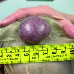
Infantile Hemangiomas (IH) are the most common benign tumor of infancy, occurring in over 10% of newborns. The head and neck is the most frequently affected area (60%), and the scalp is a typical site for such large lesions. Scalp-IHs are usually focal lesions that can be both disfiguring and may lead to complications such as ulceration and bleeding. We describe a case of a 30-months old female who presented a large scalp-IH at birth that rapidly grew in the first year of life. Topical and systemic treatments (with timolol ointment and oral propranolol, respectively) were not effective in reducing dimensions of the hemangioma. After vascular imaging study, the patient underwent surgical resection of the IH and primary closure with excellent cosmetic outcome. When medical therapy is ineffective or cosmetic and functional integrity is threatened, early surgery allows to completely removing large scalp-IHs, with good cosmetic results.
Downloads
Citations
Supporting Agencies
Infantile hemangioma; Surgical management, Magnetic Resonance Imaging Propranolol, Scalp.How to Cite

This work is licensed under a Creative Commons Attribution-NonCommercial 4.0 International License.