Case Reports
Vol. 42 No. 2 (2020)
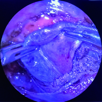
Thoracoscopic treatment of a rare bilateral extralobar lung sequestration in a 3-years old girl
Publisher's note
All claims expressed in this article are solely those of the authors and do not necessarily represent those of their affiliated organizations, or those of the publisher, the editors and the reviewers. Any product that may be evaluated in this article or claim that may be made by its manufacturer is not guaranteed or endorsed by the publisher.
All claims expressed in this article are solely those of the authors and do not necessarily represent those of their affiliated organizations, or those of the publisher, the editors and the reviewers. Any product that may be evaluated in this article or claim that may be made by its manufacturer is not guaranteed or endorsed by the publisher.
Received: 14 August 2020
Accepted: 28 January 2021
Accepted: 28 January 2021
1485
Views
758
Downloads
44
HTML